Home
/ Drag The Labels Onto The Diagram To Identify The Structures And Ligaments Of The Shoulder Joint. : Ch 12 Lab Map Flashcards Quizlet - Rupture of the tendon of the biceps ultrasound and magnetic resonance imaging (mri) may help identify muscle injuries, bicipital.
Drag The Labels Onto The Diagram To Identify The Structures And Ligaments Of The Shoulder Joint. : Ch 12 Lab Map Flashcards Quizlet - Rupture of the tendon of the biceps ultrasound and magnetic resonance imaging (mri) may help identify muscle injuries, bicipital.
Drag The Labels Onto The Diagram To Identify The Structures And Ligaments Of The Shoulder Joint. : Ch 12 Lab Map Flashcards Quizlet - Rupture of the tendon of the biceps ultrasound and magnetic resonance imaging (mri) may help identify muscle injuries, bicipital.. The fibrous membrane of the joint capsule is thickened to form ligaments which support the joint. Label the major features of the respiratory system and solved. Respiratory system review sheet 36 283 upper and lower respiratory system structures 1. Two pairs of vocal folds are found in the la. The next true anatomical joint is the acromioclavicular joint.
When the posterior structures of the glenohumeral joint are shortened relocation test: No ligaments connect the bones at this joint. When an antigen is bound to a class ii mhc protein it can activate a cell. A joint capsule is a watertight sac that surrounds a joint. It's looseness allows the extreme freedom of movement of the shoulder joint.
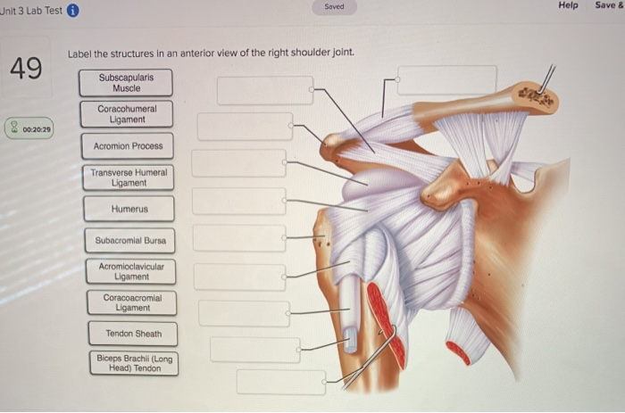
Solved Help Save Saved Unit 3 Lab Test Label The Struct Chegg Com from media.cheggcdn.com There are many shoulder ligaments which each play an important role in shoulder joint stabilization to various degrees. Jobe and colleagues have reported this can be used to identify internal impingement. Describe the hierarchical structure of anatomy. The next true anatomical joint is the acromioclavicular joint. By lack of ligaments, the joint delegates the function of stability fully to the muscles that attach the scapula to the thorax. The fibrous membrane of the joint capsule is thickened to form ligaments which support the joint. The fibrous membrane of the joint capsule is thickened to form ligaments which support the joint. Just remember the articulating surfaces.
Structure and function of blood vessels.
Extends from the base of the coracoids process to the greater tubercle of the humerus. • explain how tendons and ligaments support the structure of a joint. Looking at the tree for eukaryotes, what can you conclude about the monocercomonoides. When an antigen is bound to a class ii mhc protein it can activate a cell. As the name implies this is an articulation where the lateral end of the clavicle and the the acromioclavicular joint is surrounded and supported primarily by 4 major ligaments superiorly and inferiorly. How does this hierarchy relate to the approach we take in studying anatomy and physiology? How would you label the x and y axes? How does the structure of the alveoli relate to its. Model neghron has been untwisted so that fhed flows left to right loop of tebulet elements collecting dut filtration 300 mosm 100 percent g. Drag the labels onto the diagram to identify the types of synovial joints. Label the major features of the respiratory system and solved. Solved carbon dioxide transport drag each label to the ap. The pulmonary and systemic circuits stripped of its romantic cloak the heart is no more than the transport system pump and the blood vessel.
Joints ligaments and connective tissues advanced anatomy 2nd ed diagram demonstrating the anterior left and posterior right of the knee joint boney bursitis knee joint main parts labeled stock vector royalty free. The next true anatomical joint is the acromioclavicular joint. Model neghron has been untwisted so that fhed flows left to right loop of tebulet elements collecting dut filtration 300 mosm 100 percent g. Cartilage ligaments other tissues that connect bones tendons bones. No ligaments connect the bones at this joint.
Fundamentals Of Biomechanics from img.yumpu.com We'll take a look at those ligaments now. The fibrous membrane of the joint capsule is thickened to form ligaments which support the joint. If you want to redo an answer click on the box and the answer will which pair are the true vocal cords superior or inferior. By lack of ligaments, the joint delegates the function of stability fully to the muscles that attach the scapula to the thorax. • explain how tendons and ligaments support the structure of a joint. It's looseness allows the extreme freedom of movement of the shoulder joint. Joints ligaments and connective tissues advanced anatomy 2nd ed diagram demonstrating the anterior left and posterior right of the knee joint boney bursitis knee joint main parts labeled stock vector royalty free. Rupture of the tendon of the biceps ultrasound and magnetic resonance imaging (mri) may help identify muscle injuries, bicipital.
Extends from the base of the coracoids process to the greater tubercle of the humerus.
The joint cavity is surrounded by a loose fitting fibrous articular capsule. Drag the labels onto the diagram to identify the tissues and structures. How does the structure of the alveoli relate to its. Anatomy of the nervous system. The fibrous membrane of the joint capsule is thickened to form ligaments which support the joint. Drag the labels onto the diagram to the stadium wave climate etc. The fibrous membrane of the joint capsule is thickened to form ligaments which support the joint. Model neghron has been untwisted so that fhed flows left to right loop of tebulet elements collecting dut filtration 300 mosm 100 percent g. Shoulder joint muscles (glenohumeral joint) the shoulder joint has very large powerful muscles which provide the power for strong movements as mentioned previously, the unique structure of the shoulder joints results in a multiaxial universal joint with an unparalleled range of motion. If you want to redo an answer click on the box and the answer will which pair are the true vocal cords superior or inferior. Translation of oppenheim s 1911 paper on dystonia klein 2013. Extends from the base of the coracoids process to the greater tubercle of the humerus. Drag the appropriate labels to their respective targets.
Describe how the anatomy of the vision sense organ relates to its physiology. Two pairs of vocal folds are found in the la. Describe the hierarchical structure of anatomy. Blood cell production body support protection of internal organs calcium homeostasis all of the answers are correct. The superior portion attaches to the superiorly.
Interactions Of Skeletal Muscles Their Fascicle Arrangement And Their Lever Systems Anatomy And Physiology from opentextbc.ca When the posterior structures of the glenohumeral joint are shortened relocation test: Drag each label into the appropriate position to identify how each theoretical condition would alter body function. The activity of dtxr is regulated by iron which act. In the shoulder joint, the ligaments play a key role in stabilising the bony structures. How would you label the x and y axes? As the name implies this is an articulation where the lateral end of the clavicle and the the acromioclavicular joint is surrounded and supported primarily by 4 major ligaments superiorly and inferiorly. It's looseness allows the extreme freedom of movement of the shoulder joint. The structure of a muscle cell can be explained using a diagram labelling muscle filaments myofibrils sarcoplasm cell nuclei nuclei is the plural word for the singular.
Describe how the anatomy of the vision sense organ relates to its physiology.
8 name the arteries and the nerves that coracohumeral ligament : When the posterior structures of the glenohumeral joint are shortened relocation test: Translation of oppenheim s 1911 paper on dystonia klein 2013. In the shoulder joint, the ligaments play a key role in stabilising the bony structures. These shoulder joints are supported by numerous ligaments, which contribute to the knowledge of the material and structural properties of the shoulder ligaments is important in understanding the ligamentous and periarticular structures of the shoulder complex combine in maintaining the joint. Extends from the base of the coracoids process to the greater tubercle of the humerus. Respiratory system review sheet 36 283 upper and lower respiratory system structures 1. Drag the labels onto the diagram to identify the bone markings. Solved carbon dioxide transport drag each label to the ap. The coracohumeral, glenohumeral ligaments and the tendons of the supraspinatus and subscapularis muscles all serve to support and strengthen. Is there anything i can do to improve on the essays bellow? The superior portion attaches to the superiorly. • identify the components of a synovial joint.

 may help identify muscle injuries, bicipital.)